In our brain dissection we used a sheep brain to look deeper into the anatomy and physiology. In the first birds-eye-view picture, you can see the red pin on the brain-stem, and all the other pins in their respective areas. The brain stem we learned was very fragile. After Kian grabbed it, it became half severed, which made it hard to distinguish some parts later on.
name
|
function
|
B-stem
|
Connecting neurons
|
cerebrum
|
Voluntary movements
|
Cerebellum
|
Regulates muscle activity
|

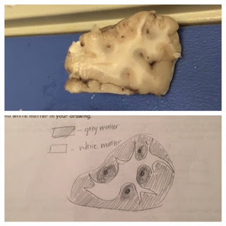
In the brain itself, it is shown clearly where the myelin is present and where it is not. Myelin wraps around axons and help the impulse travel smoothing through. In the picture right above or below, myelinated areas are whitter, compared to the darker grey that represents grey matter. After slicing the brain into two halves, we were able to identify many more structures.
Name
|
Function
|
Pons
|
Relays info from cortex and cerebrum
|
Medulla Oblongata
|
Helps with homeostasis and regulates respiration
|
Thalamus
|
Sensory, motor signal relay, and sleep
|
hypothalamus
|
Maintains homeostasis, creates essential hormones
|
Corpus collum
|
Relays information back and forth from both hemispheres
|
Optic nerve
|
Relays all the impulse to the b
|
midbrain
|
Motor control and hearing senses
|
.The brain in pictures are very "neat" and all the parts are clearly shown, but during the sheep brian dissection we found ourselves second guessing a lot besides what was grey and white matter.


No comments:
Post a Comment